3D Imaging
Not long ago, the only sure way to determine the exact location and condition of the bone underneath soft tissue was to surgically expose it. X-Rays were developed allowing us a deeper peek into the underlying bone structure of the face and mouth, but it only gave us a two dimensional view.
With the advent of Computed Tomography (CT) scans doctors now have the ability to view a complex 3-D model of your mouth, showing striking detail of the bone and tissue density, nerves, sinuses, and the exact layout of an individual’s jaw and teeth. By compiling and combining hundreds of images of the patient, this technology allows for careful planning and unparalleled precision. This results in a drastic reduction in treatment time, minimizing pain, swelling, and recovery time.
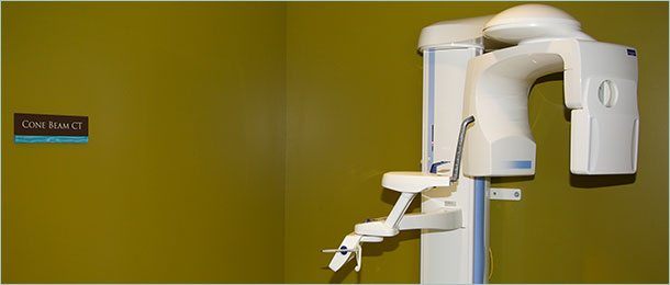
Now, what about minimizing radiation exposure? The newest generation of dental imaging comes in the form of the Planmeca Promax 3D, a state-of-the-art cone beam CT. This technology uses short bursts of radiation, instead of the continuous radiation emitted by many other scanners. The result is drastically-reduced radiation exposure for most patients, while providing accurate distortion-free 3-D images.
Tahoe Oral Surgery and Implant Center is one of the select few state-of-the-art practitioners using the Planmeca Promax 3D in both of our office locations. If you’re considering dental implants, let us apply this innovative technology, along with our expertise, to assure the best possible outcome for you and your teeth.